

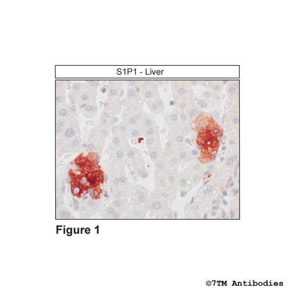
S1P1 (GP-IHC-grade), Sphingosine 1-Phosphate Receptor 1 Antibody, Guinea Pig

NEW


- Order number: 7TM0275N-ICGP
- Content: 100 µl
- Host: Guinea Pig


NEW

NEW

NEW

30
NEW

NEW
, Sphingosine 1-Phosphate Receptor 1 Antibody")
NEW

NEW
")
NEW

NEW

NEW
")
NEW

Recently viewed